Digital Inline Holographic (DIH) Microscopy
Point source digital inline holographic microscopy (DIHM) is the simplest realization of the holographic method as originally described by Dennis Gabor.
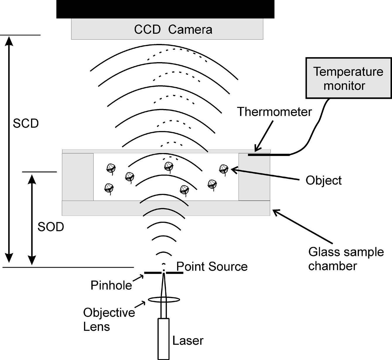 Hardware: The hardware required is extremely simple, see the schematic: A laser (L) is directed onto a pinhole (P), having a diameter of the order of the wavelength, which acts as the "point source" from which a spherical wave of wavelength l emanates. The wave illuminates an object (O), in our setup a few millimeters from the pinhole, and forms a geometrically magnified diffraction pattern on a screen (C), in our case a CCD chip, a few centimeters away. If the scattered wave, shown by dotted lines in Fig. 1, from the object is small compared with the unscattered reference wave, the interference pattern on the screen constitutes a hologram, linear in the scattered wave. The hologram is stored as a digital image in a computer.
Software: The next step is numerical reconstruction. The role of reconstruction is to obtain the three-dimensional structure of the object from the two-dimensional hologram on the screen, or, in physical terms, to reconstruct the wave front at the object. This can be achieved via a Kirchhoff-Helmholtz transform. By reconstructing the wave front on a number of planes at various distances from the source in the vicinity of the object, a three-dimensional image can be built up from a single two-dimensional hologram. For the numerical implementation of the transform we have developed a fast algorithm (patented) that employs a coordinate transformation that transforms the integral into a convolution which is solved by three consecutive Fast Fourier Transforms.
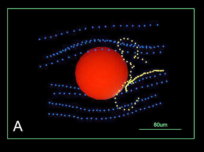
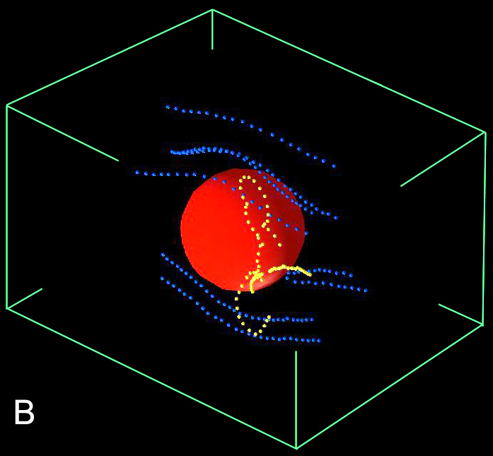
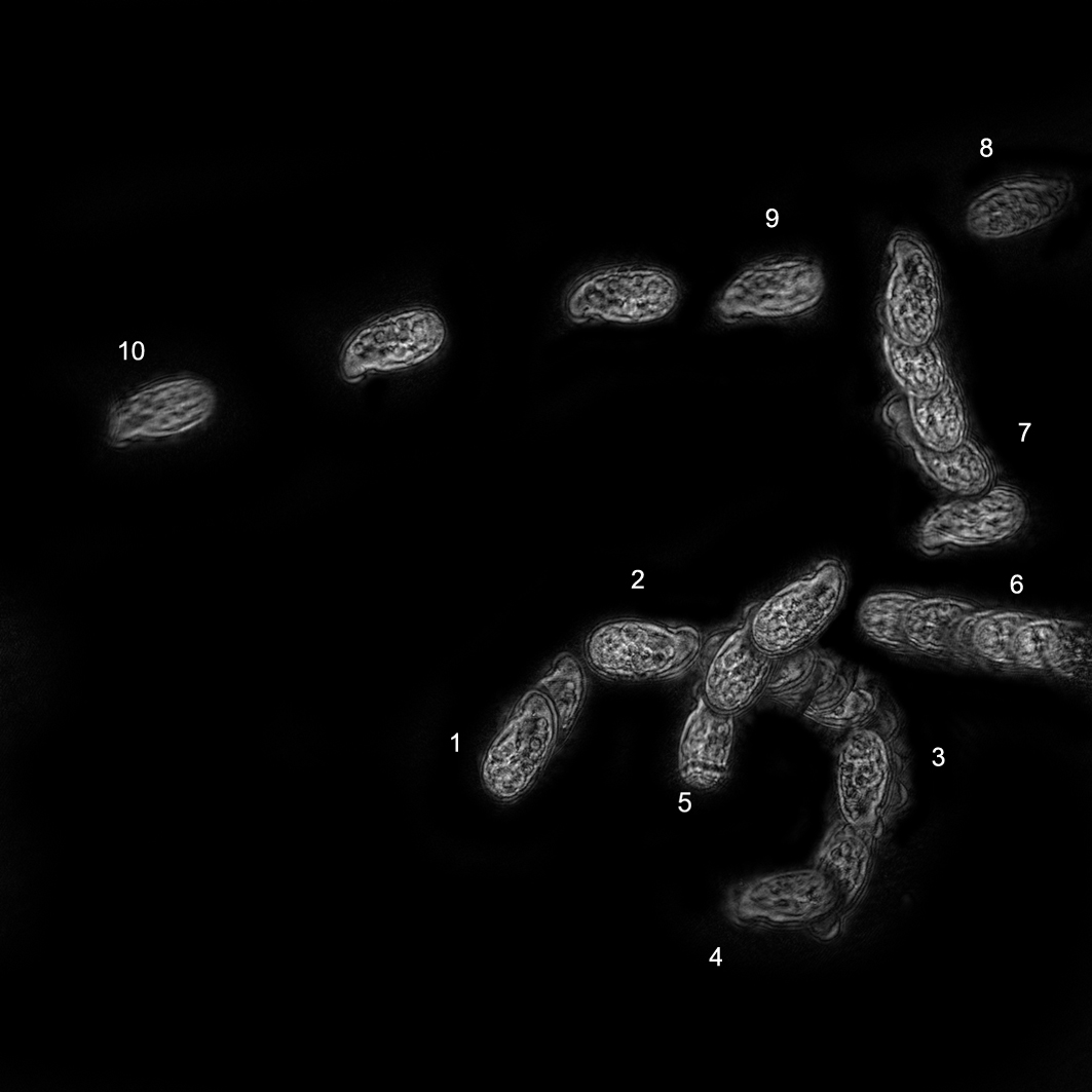
4-D Tracking: When the sample contains objects that are in motion, then high quality contrast images of these objects can be constructed if a succession of holograms is taken. The sequential position of the objects at successive recording times can be obtained by the following procedure: i) Record holograms (h1-hn) with exposure time t and at time intervals Dt. ii) Generate new holograms by subtraction of consecutive hologram pairs, i.e., (h1-h2), (h3-h4), etc. iii) Sum these difference holograms to obtain one final hologram. iv) Re-construct this final hologram to obtain images of the objects in different depth planes in the sample. This approach ensures that unwanted background effects such as interferences from sample containers and other stationary objects are eliminated so that the space and time evolution of object trajectories are clearly captured.
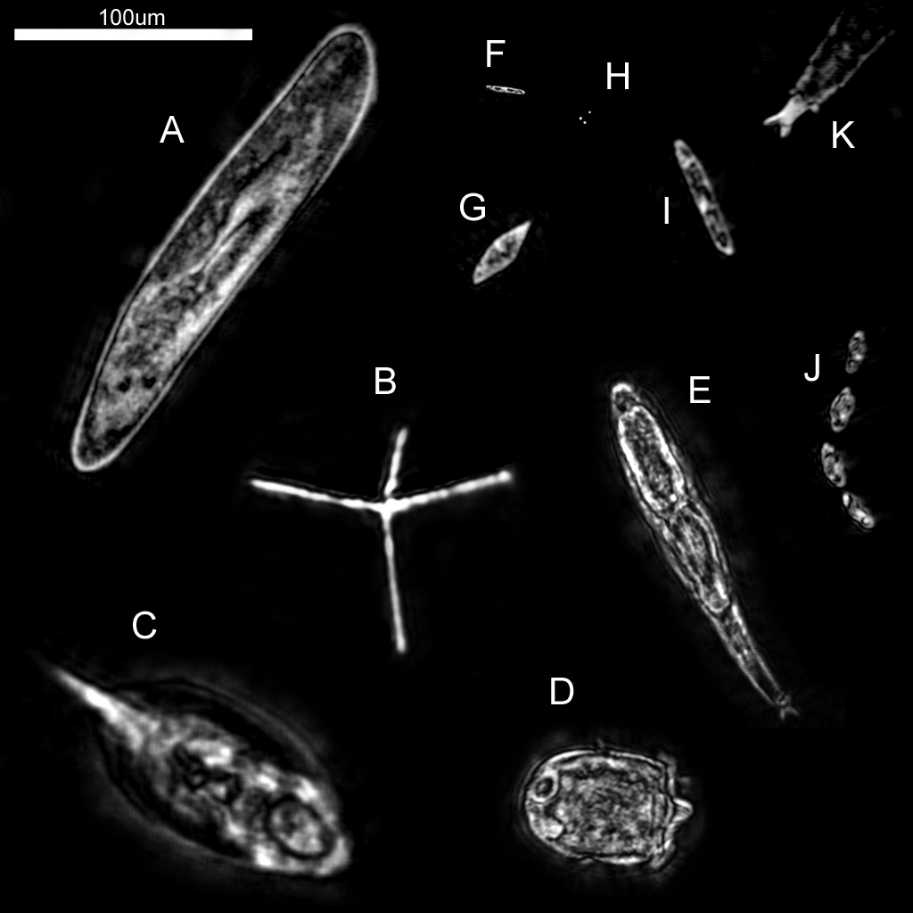
Advantages of DIH microscopy: (1) Simplicity of the microscope: In-line holography is microscopy without objective lenses. The hardware required is a laser, a pinhole and a CCD camera. (2) Speed: Changes in the specimen and 3-D motion can be followed at the capture/video rate of the CCD chip. (3) Maximum information: A single hologram contains all the information about the three-dimensional structure of the object. (4) 4-D tracking: "Adding" the frames of a holographic movie captures, in reconstruction the tracks of many objects in 3-D. (5) Maximum resolution: Optimal resolution, of the order of and less than the wavelength of the laser is obtained easily and routinely. In Figures 1-3 we give typical examples obtained with the desktop DIHM. In figure 1 we demonstrate the depth of field showing several reconstruction from one hologram of 1micron latex beads embedded in a 1 cm3 gelatin block.
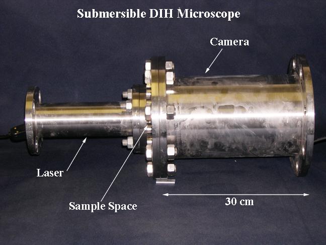
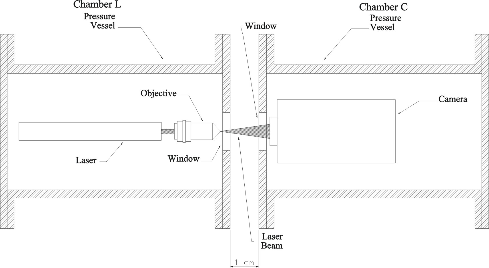
Submersible holographic microscope - SDIHM SDIHM has been designed for the remote observation of moving microscopic objects at any depth of water in the ocean, in lakes or rivers. A schematic of the design is shown in the figure together with the actual instrument. The small steel chamber on the left houses the laser and the pinhole attached to a window. The chamber on the right accommodates the CCD camera behind a second window, together with the USB-2 to ethernet converter for transmission to a boat, a buoy or a land-based station. Seawater circulates through the space between the source and camera windows so that the camera records anything moving with it, such as algae, plankton and inanimate particulates. The instrument is ideally suited for marine biology and environmental control.
DIHM - software This software package enables the automatic capture of holograms and their reconstruction. It also has many features pertinent to the processing of the images and their export to other visualization packages. The desktop DIHM instrument and the submersible SDIHM instrument as well as the DIHM - software package are available through Resolution Optics Inc (see Resolution Optics Inc. for further information).